{Internal} as opposed to the more common ecto-parasites {external}of the usual
types found on fish by hobbyists. Although Hexamita can be an ecto-parasite.
The two most common species are H Salmonis or H Truttae
and H Intestinalis. {H salmonis and H truttae
being classed by some as the same sub-species}
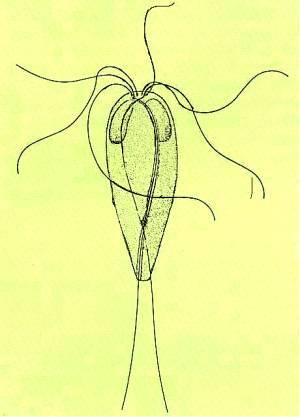
Hexamita is pyriform in shape and has two nuclei at the anterior end, it was
renamed Hexamita because it possesses 6 anterior flagella, it also has 2 posterior
flagella hence the former name of Octomita, the flagella are usually about
1.5 times the body length. With the posterior flagella just trailing where
as the anterior flagella are very motile.
Most Hexamita in koi
are found internally in the alimentary track as commensal symbionts, but
Hexamita can be found in the heart, liver, gall bladder, blood and the peritoneal
cavity. Hexamita reproduce in/on a healthy fish at a normal rate by longitudinal
binary fission and although are pyriform
in shape measuring about 6 micrometers across to 12 micrometers long,they
can change to a more spherical shape just prior to spitting.
Schizogony is actually achieved by Hexamita invading the epithelial
cells of the intestine where the nucleus combines to form a single nucleus
this single nucleus can then divide half a dozen times within one single organism.
This division is really quite rapid when you consider the normal binary fission
method of reproduction can take place in less than 24 hours at which time
many newly formed Merozoites are ready to re-infect new cells and the whole
process starts over.
Transmission
Transmission is by mouth although there is evidence Hexamita can infect by
the anal route. Scientific evidence has also confirmed the presence of Hexamita
internally in frogs and other amphibians. The encysted stage can survive for
weeks away from the host lying in faeces, so we can deduce that transmission
and infection can be very varied indeed.
Hexamita forms into a cyst to infect the host internally by the oral route,
as it cannot get past the part
of the intestine where typical stomach acids are made for digestion in fish
that do develop stomach acid {typical ph 1 to 2.5}, this would destroy them
so they are protected from this by the formation of a
cyst
Hexamita make their way to the Lumen in the upper intestine or ceca and the
free-swimming stage will live in the stomach fluids and as previously mentioned
will reproduce normally and live along side the host, provided thefish remains
healthy.
Symptoms
Flashing may occur, the fish may look emaciated with sunken abdomen, you may
be excused for thinking the fish was suffering from nutritional deficiency.
Other signs are anaemia (light coloured gills lamellae), and a yellow cast
to the faeces. In severe cases fish suffering from hexamitiasis will also
go listless and point into a corner of the pond/tank and not feed at all,
the fish will go literally off colour, {skin will darken}
If I fish dies and there are no outward signs other than the descriptions
given above squash tissue samples and blood samples should be taken ASAP and
viewed under a microscope, in a suspected live fish a fresh sample of faeces
should be prepared on a slide and examined under a microscope, this is
not possible in advanced stages of the disease as the fish will have long
stopped eating and the stomach will be empty.
Hexamita is easily seen under the scope at any magnification between x100
and x400, provided a thin sample is presented under the objective, easy enough
with a blood sample, not so easy with tissue or faeces samples, in this case
a squashed tissue or faeces sample should be spread over the slide and another
slide pressed down onto the sample and squeezed together between the thumb
and for-finger, after separating the two slides this process should be repeated
with another clean slide and if necessary repeated until a very thin sample
is obtained, then finish with a cover slip.
A good light source is a positive advantage turning the light well up and
shutting down the iris for good contrast, focusing the condenser so the light
is concentrated onto the parasite for good viewing.
Treatment
Treatment is very difficult with advanced stages of the disease The most effective
treatment is with Metronidazole Flagyl tm, in the form of 250 mg tablets available
from some vets in the UK and the US, it's used for intestinal bacterial infections
also intestinal protozoa. It is however an antibiotic, so I strongly recommend
you do not use Metronidazole in the pond/fish tank water, as this will result
in the immediate and total demise of your biological filter, its an indiscriminate
antibiotic.
As a bath, one 250mg tablet crushed and dissolved into a little warm water
then poured into 50 litres of water then submerse the fish for three days
with a raise in temperature and air stones, either water changes or filtering
water over activated carbon to rid the system of the medication
{warning
Duncan