www.koi-unleashed.co.uk
ALL THINGS KOI AND H2O
Koi Anatomy
By Phil Ishizu
& Spike Cover
Anatomy is
defined as the morphologic structure of an organism. Morphology is defined as the science concerned with the
configuration or the structure of animals and plants.
External Anatomy:
Form – Koi have what is known as a fusiform shaped body (tapering toward each end) as seen in the diagram below from Living Jewels (reproduced with the kind permission of the authors, Ronnie Watt and Servaas de Kock).
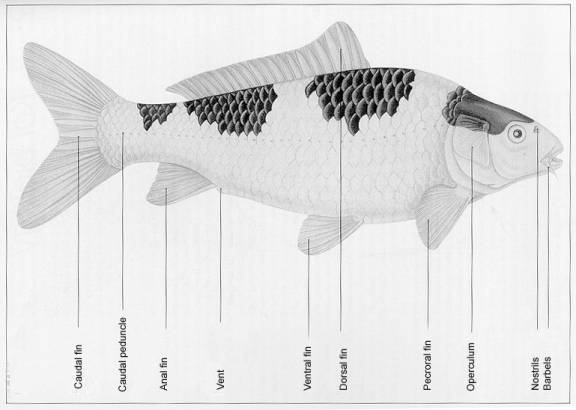
Fins – Koi have 3 single fins and two sets of paired fins.
The caudal or tail fin is primarily used for forward swimming especially fast swimming. The dorsal (top) fin is used for stabilization during forward motion. The largest leading ray becomes very stiff, sharp and thorn-like as the koi grows older.
The anal fin, like the dorsal, is used for stabilization. Also, the largest leading ray becomes very stiff, sharp and thorn-like as the koi grows older.
The pectoral fins are paired and are used for numerous functions including: steering during forward motion, slow swimming both forward and backward, breaking and to counteract the jet effect of the water being forced out of the opercular openings.
The pelvic (or ventral) fins are also paired and serve to control pitch and roll and to counter lift.
The fins are thin and well vascularized, which makes them vulnerable to damage. It is also easy to see or detect changes and damage to the fins. Therefore, diseases are often first detected in the fins, which can appear to be damaged, torn, or hemorrhaging.
Cuticle
The skin and scales are covered by the cuticle, a non-cellular mucus coating. More commonly known as the slime coat, the cuticle of the koi is a thin layer of mucus that contains many protective substances including antibodies, lysozyme (an enzyme that is destructive to cell walls of certain bacteria), and C-reactive protein (a protein that may have some antibacterial properties). The cuticle is the koi’s first line of defense against water-borne irritants and parasites and it assists the skin with drag reduction for better locomotion.
Skin
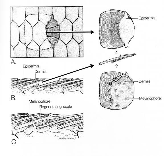
The cellular layers of the skin consists of an epidermis, dermis, and hypodermis. The epidermis (outermost cellular layer) is very thin, usually only 6 to 8 cells thick and contains unicellular mucous glands with a network of very small capillaries. The dermis (middle layer) contains the scales, the scale forming cells, pigment, blood vessels, and nerves. The hypodermis is a vascularized fatty layer between the epidermis and the muscle or bone beneath. It is the interface between the skin and the rest of the body.
The following illustration is of the skin of a swordtail (tropical livebearer) but with the exception of the outline shape of the scales (which are shown later in this section), is very similar to carp (koi) and is reproduced with the kind permission of Tetra Press from the book, Aquariology, The Science of Fish Health Management – Master Volume, edited by John Gratzek and Janice Matthews.
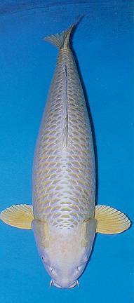
Scales – All koi scales are of the cycloid type and most koi have scales over most of their bodies but lack scales on the head. These koi scaled koi are referred to as wagoi in Japanese.
Some koi have scales only along the dorsal line and the lateral line; these are called German scaled koi (Doitsu, in Japanese). Some koi/carp are scale-less and are referred to as leather koi/carp. Other koi/carp have a heavy scales appearing almost randomly and are referred to as armored scaled koi/carp.
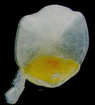
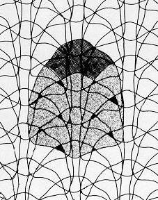
Scales are thin flexible plates with a layered structure that grow from the dermis. Note that since the scales are produced in the dermal layer, removing a scale creates an ulcer, or hole in the skin, which can be a potential area for pathogens to enter the body. The scales grow essentially from the center outward. The actual origin of growth is in the center of the scale and is seen in the scale below (courtesy of Mark Whalen): Note Scales are normally flat, however the picture of the scale below had been off the fish and dried for one to two years prior to scanning.
A detailed look at the growth rings as seen through a microscope appears like the following at 100x (courtesy of Brett Rowley)
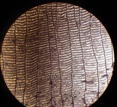
Note: the age of a koi cannot be determined by simply counting the rings on a scale (as can be done with a tree from a cross sections of the trunk).
There is a tremendous amout of overlapping in scales and there can be up to six or seven layers in some spots (small dark areas). About 20% of most scales are exposed to the exterior, i.e., the portion of the scale without overlapping scales, see large dark area of illustration above (provided by Masaki Okamoto).
Note: On some koi, the dermis grows from beneath the scale and is seen as ‘fukurin.’ Varieties such as ogon and asagi typically display fukurin especially on the shoulder area. It appears around the external edges of the scales.
On the main body of the fish shown on the platinum or white areas are the fukurin while the darker or yellowish areas are the scales.
Mouth
Koi have a protrusive mouth that is ideally suited for bottom feeding. Koi are referred to as benthic feeders, since the position of the mouth on the underside of the head allows them to literally suck foods from the pond bottom. However, they are able to feed at any level, bottom, mid-water and at the surface.
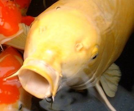
Koi have no teeth in their jaws and this is consistent with how koi in nature feed; i.e., suck everything up, taste it, and then decide if it is consumable or not. The Koi do however, have teeth like structures (pharyngeal teeth) on the pharyngeal bones located just behind the gill chambers. Food is ground by these teeth against a bony pad (carp stone) located on the top of the pharynx, itself.
Sensory Organs
Hearing:
Koi do not have external ears but hear by an internal “ear” that is connected via a group of bones, known as the Weberian ossicles to the cranial swim bladder. It is believed that sound is amplified by the swim bladder. Koi, like other fish, are very sensitive to sound, and can be stressed to the point of becoming ill when exposed to loud noises on a constantly recurring basis.
Eyes:
Koi eyes are similar in structure to our
own. However, unlike ours, they have bilaterally placed eyes that are
independently movable, which increases the range of area in their visual
field. They have cones and rods, the
structures of the eye that see color and black and white, respectively. They
probably have good enough sight to see the shape of words on a printed page.
Since koi live in a water environment, they do not need protective
eyelids. This, however, makes their
eyes more vulnerable to abrasion or other mechanical trauma during netting and
handling.
The lenses are spherical shaped and protrude through the pupil which allows considerable peripheral vision, including forward and rear. In healthy fish, the lens is completely clear. Bacterial and nutritional problems often will cause the lens to become cloudy.
The pupils are under neurological control like mammals, but the response time is too slow to be of clinical use.
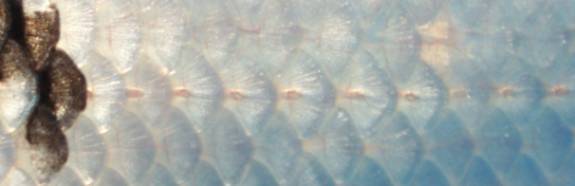
Lateral Line
The lateral line runs along the side of the koi about midway down the side of the fish. Holes in the scales lead to a canal beneath the surface that contains neuromast cells. Water movement in any direction striking the sides of the fish will cause the mucous in the canal to vibrate. These vibrations stimulate the neuromast cells that are linked to the periphery nerve system and provide one the most effective perceptions for survival (flight reaction).
The lateral line is an important landmark. It is at approximately the same level as the spine, which has a blood vessel that runs along the length of the spine just ventral (underneath) to the spine and will be important for locating the blood vessel.
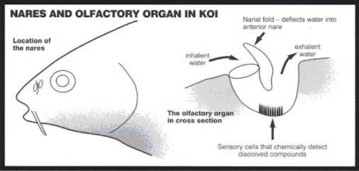
Olfaction
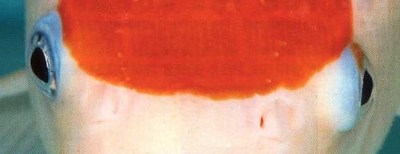
The olfactory organs (used to smell) are located at the base of the nostrils called nares. Water does not flow to any other part of the body from the nares. They are exclusively used for olfaction (smelling), are paired and located between the eyes and the mouth. They are shaped like and can be thought of as small U-tubes into which water enters through the leading or forward hole and exits through the rear port or opening. Just behind the forward opening there is a flap of skin that directs water into the forward opening of the nare as the koi moves forward in the water. The movement of substances through the nares is aided by diffusion and by the motion of small hairs-like structures (cilia) within the nares. The following illustration is slightly modified and reprinted with the kind permission of Koi Carp magazine.
Taste:
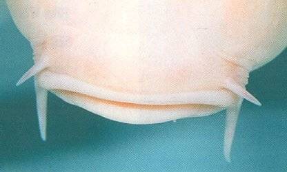
Taste buds are numerous in and around the lips, mouth and tips of the barbels. Koi have two pairs of barbels. Three hundred years ago they had three pairs.
Internal Anatomy
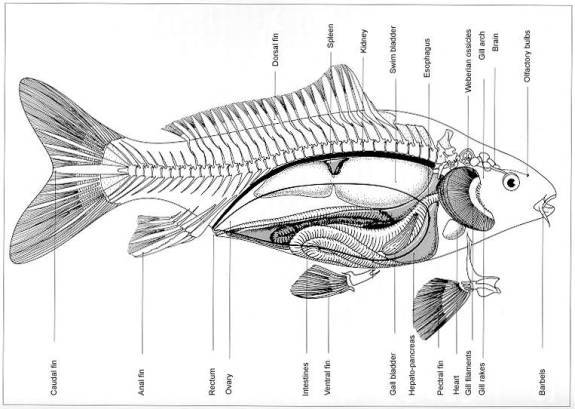
The internal organs are placed
approximately as shown in the illustration below (reproduced with the kind
permission of the authors of Living Jewels).
The first portion of the gut is elastic and can be used to store
food. It is generally accepted that koi
do not have a stomach (wide hollow organ that functions to mix food with mucus,
acid and digestive enzymes).
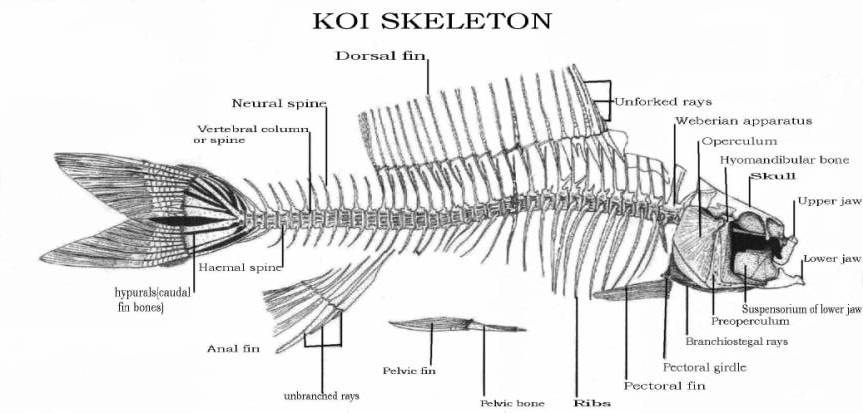
Bones
Carp are Teleost fish which means
literally “bony skeleton” and carp are one of the boniest fresh water fishes. Fish bones are thin and light weight with no bone
marrow in the center. A light weight skeleton is advantageous to an animals
that needs to be buoyant to live in a water environment.
The drawing below is taken from the Tetra Encyclopedia of Koi and reproduced with the kind permission of Interpet Publishing Ltd. It was redrawn from the book by Duncan Griffiths.
Muscle
Generally two types of muscle exits in Teleost fish, white and dark muscle. The white muscle is thought to be for fast quick swimming and the dark muscle, located directly under the skin if for sustained swimming. The clinical significance of these different types of muscle is that there is much different drug kinetics when injections are made into them. It is generally accepted that red muscle is well vascularized (has a good blood supply), which would facilitate absorption of medicines into the blood faster than white muscle, and sustain the release since the blood flow is likewise sustained in comparison to white muscle which can create an oxygen debt and requires a recovery period.
Gills
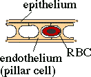
The gills are a complex system made up of both bony and cartilagnious stiffened arches. Each side of the fish has four gill arches. On the exterior side of the gill arch are the gill filaments, which are made up of lamallae and secondary lamellae structures, the latter being only 2µ thick. It is these structures that are responsible for the exchange of gases (O2/CO2) through the epithelial cells. The epithelial cells are the direct link between the O2 in the water and the fish’s blood stream.
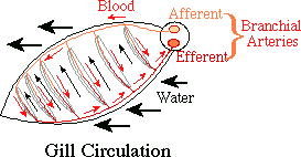
The above illustrations were reproduced with the kind permission of Paul Maslin and are from his web site at: http://www.csuchico.edu/~pmaslin/ichthy/fshrsp.html
The gills also have specialized mucus-producing cells, and chloride cells which function in mucus production and osmoregulation, respectively.
Covering the gill arches on the outside of the fish is the operculum or gill cover. This is a multi-purpose part of the koi’s anatomy with the primary function being to help control the pressure of the water taken in through the mouth and passed along the gill filaments.
Also found on the gill arches, but more to the inside or anterior part of the arch, are the gill rakers which act as food filters. Clogged rakers are often the cause of flashing or head shaking as the fish attempts to clear the gills of excess food after eating.
Circulatory System
The heart is the pump that moves the blood through the system. It is located low in the fish between the gills. A koi's heart is a two-chamber organ having a ventricle and an atrium. Although it has been described as a four-chambered organ, the extra two chambers, before and after the main pumping chambers, are called the sinus venosus and bulbus arteriosus, respectively. They are smaller than the main pumping chambers, functioning as accumulators (to smooth out any pressure surges and protect the cardiovascular system from overpressure) and, as such, are not equipped for pumping blood. They have no muscled walls but are elastic similar to balloons. The blood pressure of fish is considerably lower than that of mammals.
Blood flows from the heart through the gills and then is distributed to the rest of the body. Blood is collected into veins that eventually return to the sinus venosus just prior to returning to the heart. Fish, unlike mammals, have only one circulatory pattern. Mammals have a systemic circulation and a pulmonary circulation. One of the important veins, the caudal vein, is located ventral (under) the spine. This vein is the most accessible in the fish for obtaining blood samples.
Normally blood flows from an artery to an arteriole to a capillary to a venule into a vein and then back to the heart. A portal system is one in which the blood flows from a vein into a capillary and then back to a vein on its way back to the heart. Fish and mammals have a portal circulation of the liver. That is, blood from the intestines flows into capillaries in the liver before collecting in the hepatic portal vein.
Fish also have a renal portal system. Carp (koi) have a system that is somewhat modified from the norm. In carp the blood returning via the caudal and segmental veins is divided into the renal portal vein and a shunt to the intestinal vein (see diagram below)
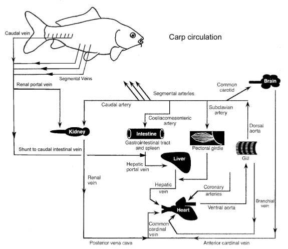
Kidneys
There are two kidneys in a koi. The caudal kidney is long and narrow, running nearly the length of the body cavity and located just below the spine. The other is the anterior, head or cranial kidney. It is located just above the heart and also contains Thyroid follicles.
Liver
The liver is smooth, dark red-brown color and is next to the cranial portion of the intestine. The right lobe of the liver covers the gallbladder and the left lobe encases the spleen.
Swim bladder
This is a two-chambered organ located directly below the kidney that is directly below the spine. There is a small connection between the two chambers that also connects to the gut. The caudal-most chamber is relatively inflexible but the other chamber has some flexibility.
Gonads
The paired gonads are located between the swim bladder and intestines. They can be separate or fused. Testes are white and fissured and ovaries are pink and smooth. The gonadal pore has a separate opening from the waste pores at the anal vent. Gonads enlarge during breeding season to almost 70% or the body weight in females and about 30% in males. Some koi can have both male and female gonadal tissues. These hemaphrodites have external characteristics between a male and female.
Acknowledgements:
This section was started by Phil Ishizu, completed by Spike Cover and was reviewed and made better by the comments and contributions of Sandra Yosha, D.V.M, Duncan Griffiths and Richard E. Carlson.
Bibliography
Fish Medicine – Stoskopf, Michael, W.B. Saunders Company, Philadelphia, PA
Aquariology, Master Volume – Gratzek, John, Tetra Press, Blacksburg, VA
The Tetra Encyclopedia of Koi – Tetra Press, Blacksburg, VA
Living Jewels – Watt, Ronnie; de Kock, Servaas – Delta Books, Johannesburg, S.A.
Koi Carp – Freestyle Publications, Poole, U.K.
